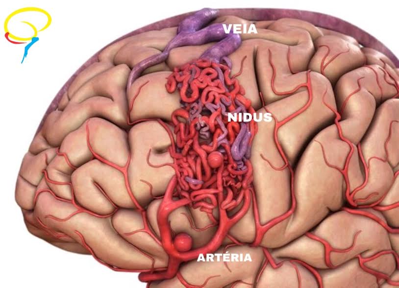
Arteriovenous malformations are abnormal connections between cerebral arteries and veins, with no capillary bed between them (small microvessels that nourish the brain), forming a tangle of blood vessels called nidus.
Due to the lack of the capillaries, blood flows quickly and directly from the arteries to the veins, bypassing the surrounding tissues. This condition causes the following symptoms:
Cerebral hemorrhage / Stroke (40-50%);
Seizures (30%);
Neurological symptoms (muscle/motor weakness, sensory impairment, speech difficulty, etc.);
No symptoms (15% – Occasional finding);
In this context, it is noteworthy that the cumulative risk of an AVM rupture and
intracranial hemorrhage is 2.3% per year.
An AVM diagnosis is established by magnetic resonance imaging of the brain, and cerebral angiography (gold standard).
Currently, there are three different surgical options for treating AVMs::
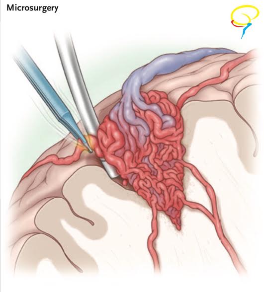
Microsurgical resection: the AVM is resected and the arteries and veins that used to fill it with blood are closed.
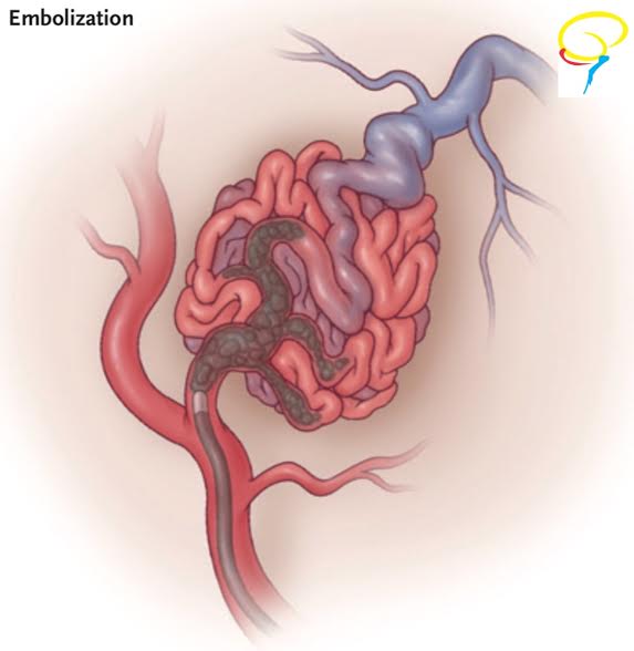
Endovascular embolization: using small catheters inserted into your blood vessels, glue or other obstructive materials is inserted into the AVM so that blood no longer flows through the malformation
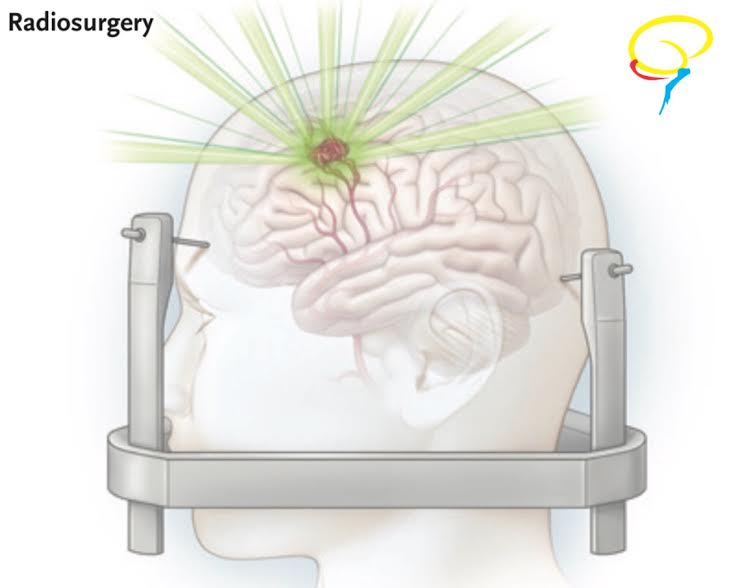
Radiosurgery: Highly targeted radiation beams are directed at the AVM to damage the blood vessels and cause scarring.